


Controlling blood glucose levels within a normal range is the objective of diabetes management and will help to prevent long-term complications such as damages to the eye, kidneys and the nervous system and may also reduce the risk of cardiovascular events such as a heart attack or stroke. To achieve good glucose control in a safe manner without hypo- or hyperglycaemia, many patients with diabetes need to check their blood glucose level several times per day before making a treatment decision. The standard self-measurement requires patients to perform a finger prick with a lancet needle and measure the glucose content of the obtained capillary blood drop with a handheld glucose meter system. A patient diagnosed with diabetes type 1 at age 20 may easily have to perform more than 75.000 of these painful finger pricks during a lifetime, each causing a small injury to the skin.
The holy grail of glucose monitoring
A noninvasive way to measure or estimate blood glucose levels without any pain and injury is therefore generally regarded as the holy grail of glucose monitoring. Fuelled by the strong patient need and the prospect of commercial success of a noninvasive monitor, many scientists and companies have entered this field of research in the last decades. A wide array of measurement techniques, including spectroscopic, optical rotation, fluorescence and electrical impedance measurements have been developed to directly or indirectly track glucose levels through the surface of skin, in the eye, in breath and most notably in bodily fluids that are readily accessible such as sweat, saliva and tears. Of all the approaches and techniques that have been tested to date, only one development has resulted in a commercial product, the GlucoWatch® G2 Biographer (Cygnus, Redwood City, CA). This wrist-worn monitor was available in selected countries in the early 2000’s for a short time period only. Selling of the device was stopped because of multiple issues: (1) the measurement wasn’t as pain- and injury-free as many had hoped, the reverse iontophoresis method, a low electric current actively pulling glucose through the skin, could result in local skin irritation; (2) the measurements were not always accurate and (3) to get readings there was still a need to perform finger pricks for multiple calibrations from a blood glucose meter. These issues highlight some of the difficulties commonly associated with noninvasive monitoring approaches.
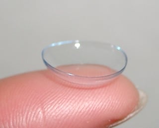
Smart contact lens project
When in January 2014 Google announced to be working on a smart contact lens to measure glucose in the tear fluid of the eye, many (including me) hoped and perhaps even believed that a working noninvasive glucose monitor would soon become reality. The image of a prototype contact lens with embedded sensor and electronics that was included in the announcement at least gave the impression that this project was well passed its initial technical development stage. Now, almost 3,5 years later, it seems that plans to start clinical testing in 2016 were postponed and the initial enthusiasm for a quick fix of needle-free glucose monitoring has passed. Should we really start worrying that this technology will fail to produce a reliable alternative to blood glucose monitoring?
Not according to the companies now responsible for carrying the project forward, Verily (a biotech spin-off formed by Google) and Alcon (eye care division of Novartis) seem confident with the project’s progress based on the information on their website or from news reports. Another reason not to worry could be that the process of elevated expectations followed by a period of disillusionment is a natural phenomenon for new technologies, called the hype cycle, although this by no means is a guarantee for success in the long run.
Tear glucose ≠ blood glucose
To better understand whether a glucose sensing contact lens or any other noninvasive monitoring of glucose in tear fluid is feasible, one needs to understand the physiology of glucose in tear fluid. Because even when miniature glucose sensors can be produced that accurately measure glucose in minute quantities, a prerequisite for a working device is that the glucose concentration in tear fluid can be used as a reliable indicator for the actual glucose concentration in blood. A valuable source is a review published by Baca et al. in 2007 providing an extensive overview of the physiology and measurement of glucose in tears from the 1930s onward. The article presents that various attempts to measure tear glucose concentration and to correlate results with blood glucose concentrations have provided contrasting and frankly unconvincing results. Various confounding factors apart from an obvious physiological time lag between blood and tear glucose that may influence the glucose concentration in tear fluid, independent of blood glucose variations, are identified:
- Implausible high concentrations of glucose have been measured in tear fluid after (i) mechanical stimulation of the eye, e.g. through rubbing, an effect that can last up to 60 minutes. It is believed that mechanical irritation of the conjunctiva (the cells lining the inner part of the eyelids) results in a leakage of glucose from epithelial cells or interstitial fluid directly into the tear fluid covering the eye. Peak tear glucose concentrations have also been observed after (ii) eye contact with water, e.g. when washing the face or during swimming. The higher concentrations are probably due to a local osmotic effect, and an additional effect through eye rubbing in response to water in eyes seems also plausible.
- Implausible low concentrations of glucose have been measured in tear fluid after stimulation of tear production as response to emotional, chemical (e.g. the smell of raw onion) or physical (e.g. bright sunlight, dust particle, physical activity) stimuli. The reason is that stimulated tears in comparison to the continuously produced basal tear fluid have a different composition and lack almost any glucose. Stimulated tears are derived from the main lacrimal glands, whereas a substantial part of basal tear fluid is derived from smaller glands in the conjunctiva which are believed to be the main source for glucose in the tear fluid.
- Not only may non-physiologically high or low readings impair the reliability of a tear glucose monitor, its measurement variability may also be substantial. Large differences sometimes exist between measurements of tear glucose in the left and right eye of the same individual. It is unclear whether these are systematic differences between the two eyes or whether the differences vary in response to various stimuli. In the first case, the observed differences do highlight the need for patient-specific calibrations for tear glucose; in the latter case, redundant glucose sensing using both eyes could be an option to filter out non-physiological glucose responses observed in one eye only.
Ambient factors
In my opinion the above identified issues are largely due to the unprotected environment of tear fluid. Blood and interstitial fluid are well isolated from ambient factors and can thus be used as a reliable sample medium. In contrast to blood and interstitial fluid no evidence exists for a function of glucose in tear fluid, and it seems that it’s not in the body’s interest to actively control the glucose content in tear fluid. Tear fluid is constantly exposed to ambient factors such as temperature, humidity and wind. Changes in these ambient factors will affect the tear fluid layer of the eye and consequently its glucose content. Cycling on a cold winter’s morning will make your eyes fill up with tears. Stepping into an air-conditioned room may have a substantial effect on tear fluid evaporation rate.
The idea of embedding sensing technology in a contact lens could be a great idea if the lens would work as a protective layer that eliminates the potential confounding effect of the ambient factors (some of you may have noticed that when cutting onion while wearing contacts your eyes don’t start swelling with tears). However, many other stimuli and responses, like emotional stimuli, activation of sensory nerves in the nose or mechanical stimulation of the eye (e.g. daily lens placement and eye rubbing) cannot be controlled by a lens and can potentially still alter tear glucose concentration. A correlation between blood and tear glucose in contact lens wearers has not yet been established.
Tear glucose and diabetes
A last major challenge for a noninvasive tear glucose monitor is the target population: patients with diabetes. The health condition of the eye and the function of tissues contributing to tear production are generally affected by diabetes which may alter the physiologic tear composition and could lead to large inter-individual differences between patients. Retinopathy and dry eyes are for example more common in patients with diabetes. Because of impaired eye health with diabetes, concerns regarding contact lens wear in patients with diabetes have been uttered, but contact-lens induced complication rates seem to be similar between diabetes and non-diabetes patients.
In summary, apart from the technological challenges to noninvasively measure tear glucose, the physiological tear glucose content seems to be easily affected by multiple stimuli and presents probably the largest hurdle in the development of a monitoring device. The first clinical results from the Verily/Alcon smart lens program are highly anticipated to provide further insight into the feasibility of using tears for noninvasive glucose monitoring.