


Why FMD?
Endothelial dysfunction plays a pivotal role in the pathogenesis of progression of atherosclerotic disease. While in the past invasive cardiac catheterization in the coronary arteries was used for the quantification of endothelial function, a non-invasive technique would of course be much more appealing. Flow-mediated (vaso)dilation (FMD) of the brachial artery is such a technique and has shown prognostic value when it comes to cardiovascular (CV) disease. Several meta-analyses have found that there is a significant inverse association between FMD and future CV events: A 1% decrease in FMD was associated with 13% increase in risk of future CV events and similarly a decrease of 1% standard deviation caused an increase of CV event risk of 22 %. FMD has proven to be a powerful research tool as well as a tool for risk stratification with a prognostic value that is difficult to ignore.
The Physiology Behind FMD
The vascular endothelium, a blood vessel’s innermost lining, is made up of endothelial cells. This monolayer regulates the tone of a vessel which affects its blood flow. The ability of the vessel to increase its diameter, leading to increased blood flow (in accordance with the Hagen-Poiseuille equation), is what is being measured during FMD. This function is vital for the homeostasis of the vessel. Other functions of the endothelium include immune response and cell adhesion. The nitric oxide (NO) synthesis in the endothelium is key for vascular homeostasis. NO, a product of the endothelial nitric oxide synthase, is released upon certain triggers, e.g., shear stress or acetylcholine. NO then diffuses to the smooth muscle cell causing the muscle cell to relax (and subsequently the vessel to dilate). Therefore, FMD depends on the formation of NO upon shear stress as well as its inactivation and NO sensitivity of the smooth muscle cell. On the scale of things, FMD measures endothelial function.
Endothelial Function Assessment
FMD measures the percentage diameter change in the brachial artery after inducing reactive hyperemia. The set-up for measuring FMD is fairly simple (Figure 1). The procedure at Profil is as follows: in short, after at least 10 minutes of rest in supine position, the arm of the subject (after fasting with no caffeine intake for at least 12 hours) will be scanned using a high-resolution ultrasound scanner with a 12.0-MHz linear array transducer. After acquiring baseline measurements of the brachial artery at rest, a cuff will be inflated over the forearm to > 200 mmHg for 5 minutes. Then, the cuff will be released and the lumen of the vessel will be measured for the subsequent 2 minutes. The measurements will then be analyzed using automated software (Figure 2). The software measures the media-media distance from a longitudinal view along the axis of the brachial artery at baseline and during reactive hyperemia. FMD is then calculated with the following equation:

Subject preparation as well as precise assessment day exclusion criteria and strict lab conditions (e.g. a temperature controlled room, no direct sunlight and no noise nor other means of distraction) may help to improve quality of FMD assessment and are implemented in Profil’s set-up for FMD assessment.
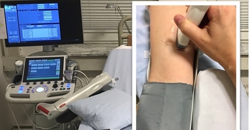 Figure 1: Set up for the assessment of FMD
Who and Where?
This method has quickly found widespread adoption, however, the learning curve to generate valid, reproducible data is long and standardization of the technique as well as measurements performed by highly skilled, experienced examiners (minimum of 100 scans per examiner) are key to high-quality results. At Profil, we have set up a rigid training program consisting of ultrasound and software analysis training that examiners must undergo prior to receiving clearance for assessment of FMD in clinical trials. A small core team conducts the procedures associated with FMD and we ensure that the same examiner performs the measurements for the same subject reducing intra-observer variability with all examiners having a proven low inter-observer variability.
Conclusion
FMD enables us to predict the risk of future CV events and is a powerful research tool when carried out with precision and care. Beside the necessary equipment and lab conditions, the examiner’s skills are vital for this method. Profil has prepared a online seminar for you in which we will discuss the key elements of FMD assessment as well as present our FMD lab to the public.
Make sure that you do not miss the online seminar on FMD and see Profil’s FMD lab for yourself.
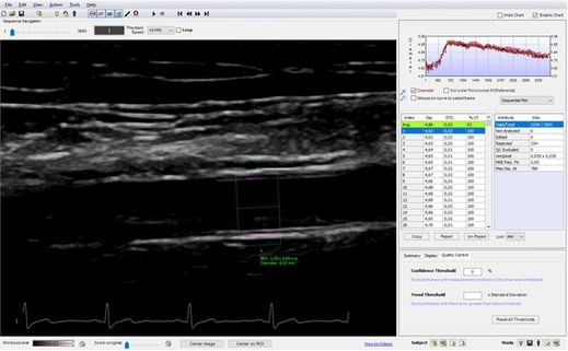
Figure 2: Automated FMD analysis of a hyperemia loop using Brachial Analyzer, Medical Imaging Applications, LLC